
Bella Shmaltsuyeva, HT (ASCP), QIHC
Senior Research Technologist
312.503.4705 | b-shmaltsuyev@northwestern.edu
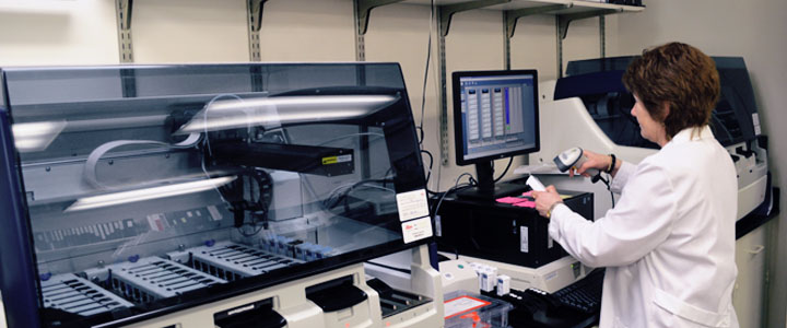
Skillful technical personnel can perform immunostaining on single tissue sections or TMA sections prepared by our histology laboratory or provided by the investigator. We offer a list of standardized immunostains and the opportunity to use other antibodies commercially available or antibodies newly developed by the investigator. The PCF is equipped with four state-of-the-art automated stainers that allow for large volume reproducible staining.
Once the tissue sections are prepared by histology, slides are sent to our IHC Lab for further staining. The tissue on slides are then exposed to an antigen retrieval to break the covalent bonds formed by formalin fixation thus allowing the antibodies to combine with the antigens on or in the cells. Antibodies can be expressed on the cell membrane, cytoplasm or in the nucleus. Use of specific detection kits (Avidin-biotin-peroxidase or avidin-biotin alkaline phosphatase detection systems are offered) triggers chemical reactions within the tissue and ultimately allow the expression of the antibody in the tissue. The customization is done after consultation with the investigator to set up the optimal conditions for a successful result.
Pathology Core Facility
of the Robert H. Lurie Comprehensive Cancer Center
Northwestern University
Olson Pavillion, 710 North Fairbanks Court, Chicago
8th Floor, Room 8-419
312.908.5546
FAX: 312.503.1148