
Demirkan Gürsel, MSc, PhD
Scientific Director
Lab: 312.503.0324
demirkan.gursel@northwestern.edu
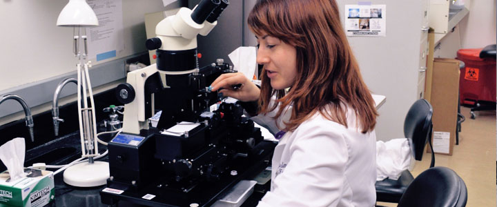
Tissue microarray (TMA) technology allows for the efficient analysis of tissue from many samples. Different size diameter cores of tissue from over one hundred tissue samples can be placed into an array and slide-based assays can be performed on this single block. This is powerful, state-of-the-art technology that allows evaluation of many tissues on a single slide while minimizing issues of variability.
TMA procedures at PCF can be done on the semi-automatic Veridiam Tissue Microarrayer VTA-100 or the Manual Tissue Arrayer by Beecher Instruments MTA-1 to create tissue microarray blocks. TMA is a technology which facilitates research by permitting the collection of biological specimens (primarily tissues) representing many patients without requiring a great deal of storage space. TMA facilities comparison of the histological characteristics of different patients' pathologies by placing many different cores of tissue together on the same TMA block, and then sectioning for microscopic slide evaluation. The TMA block, depending of the diameter size, can have in it up to couple hundreds cores. A corresponding map records each cores location in the grid and the information connected with it in an Excel sheet. The array sections can be used for all histological staining, including Hematoxylin & Eosin (H&E), Immunohistochemistry (IHC), and In situ hybridization (ISH).
See also Histology Lab.
| TMA-1 | GI | Esophagus, Stomach, Small bowel, Colon, Pancreas, Liver, Gall bladder and Appendix |
|---|---|---|
| TMA-2 | Women Reproductive | Breast, Uterus, Ovary, Cervix, Placenta, and Fallopian tube |
| TMA-3 | Male Reproductive | Prostate, Kidney, Testis, Adrenal, and Urinary Bladder |
| TMA-4 | Systemic Organs | Lung, Skeletal Muscle, Heart, Skin, Thyroid, Parotid, Thymus, Tonsil, Lymph Node |
| TMA-5 | Systemic Organs | Skeletal Muscle, Brain Cortex, Skin, Fat, Brain Cerebellum, Cartilage, Bone, Nerve, Smooth Muscle |
| TMA-1 | Normal Breast | 10 different cases |
|---|---|---|
| Ductal carcinoma | 10 different cases | |
| Lobular carcinoma | 10 different cases | |
| TMA-2 | Normal prostate | 10 different cases |
| High grade Tumor Prostate | 10 different cases | |
| Low grade Tumor Prostate | 10 different cases | |
| TMA-3 | Normal Colon | 10 different cases |
| Tumor Colon | 10 different cases | |
| Normal Pancreas | 10 different cases | |
| Tumor Pancreas | (here we have only 5 since oct 2014) | |
| TMA-4 | Normal Endometrium | 10 different cases |
| Tumor Endometrium | 10 different cases | |
| Normal Fallopian Tube | 10 different cases | |
| High Grade Serous Ovary | 10 different cases | |
| TMA-5 | Normal Lung | 10 different cases |
| Adenocarcinoma Lung | 10 different cases | |
| Squamous Lung | 10 different cases | |
| TMA- 6 | Brain: Meningioma | 2 cases, Neuroblastoma 1 cases, Glioblastoma 2 case, |
| Brain: Astrocytoma | 2 cases, Oligoastrocytoma 2cases, Anaplastic Astrocytoma 2 cases | |
| Brain: Schwannoma | 1 case, Cerebellum 2 cases, Cortex 2 cases, Pituitary 1 case | |
| Brain: Adenoca | Breast 1 case, NeuroEndocrine Colon 1 case, Mening. Anaplastic 1 case |
Pathology Core Facility
of the Robert H. Lurie Comprehensive Cancer Center
Northwestern University
Olson Pavillion, 710 North Fairbanks Court, Chicago
8th Floor, Room 8-419
312.908.5546
FAX: 312.503.1148